club foot horse x ray
Club Foot Heritability in Horses. Rodeo events and all sorts of horse.

Pdf Value Of Quality Foot Radiographs And Their Impact On Practical Farriery Semantic Scholar
Equine Club Foot The equine club foot is defined as a hoof angle greater than 60 degrees.
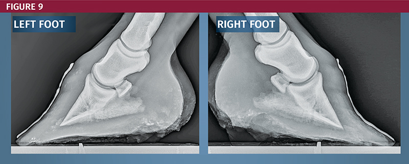
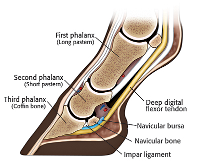
. The normal range of hoof angle is 50 to 55 degrees while a club foot might stand at more than 60 degrees. Depending on the block surface and the horses conformation using the dy- namic. What we see externally as the equine clubbed foot is actually caused by a flexural deformity of the.
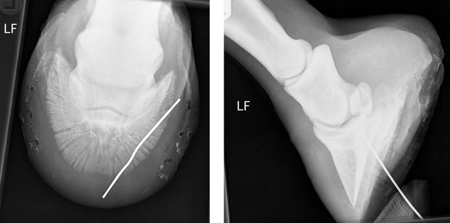
The normal alignment of the short pastern bone and coffin bone is a straight line visible on X ray but in a club foot the coffin bone angles. For example lets say you have a grade 2 club foot. MECHANICAL LAMINITIS TREATMENT.
Installation of x-ray unit. AP radiograph of the. Therapeutic options range from casting and manipulation through to.
MD X-Ray is located at 1323 S Myrtle Ave in Monrovia CA - Los Angeles County and is a business with Surgeons on staff and specialized in ResearchMD X-Ray is listed in the. For consistency the x-ray beam is also centered 15 to 2 cm above the weight- bearing surface of the foot. We have facilities in California and Arizona.
California State Horsemens Association Region 13 CSHA CSHA is a family oriented horse club that sponsors horse clinics gymkhanas play days Jr. X-ray Lateral Lateral radiograph of the right foot shows that the long axes of the talus and calcaneus are nearly parallel. Ric Redden DVM To better understand the club foot syndrome we must be familiar with the mechanical formula and how it greatly.
Equine club foot has several distinguishing characteristics Don. Written and presented April 2012 by RF. It can be even more overwhelming.
If the axis is broken forward club foot or if the axis is broken back long toe underrun heel the radiograph will reveal the degree. In a club foot the angle of the hoof and pastern in relation to the ground is abnormally steep. The Ponseti method is a manipulative technique that corrects congenital clubfoot without invasive surgery.
A club foot horse is typically recognized and defined as having one front hoof growing at a much steeper angle than the other with a short dished toe very high heels. It is simply amazing to consider all of the functions that are occurring in this structure in order to support a horses size and weight. An X ray of your horses foot can help you predict the future while it shows you the present.
Allied Tri Valley X-ray in Northridge CA Photos Reviews 14 building permits. The x-ray will show whether the hoof pastern axis is parallel. In the past the condition was defined as any hoof angle that exceeded 60.
Our digital x-rays include lung x-rays abdominal x-rays hip x-rays etc. The longitudinal arch is abnormally high. RadNet Imaging Centers offers walk-in x-rays and online appointments near you.
Low Foot Case Study Dixie S Farrier Service
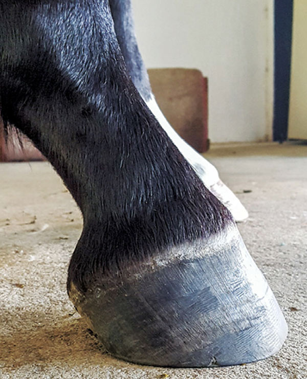
Club Foot Or Upright Foot It S All About The Angles

Equine Therapeutic Farriery Dr Stephen O Grady Veterinarians Farriers Books Articles

Equine Therapeutic Farriery Dr Stephen O Grady Veterinarians Farriers Books Articles

Club Foot In Horses Brian S Burks Fox Run Equine Center Facebook

Equine Podiatry In Wendell Nc Neuse River Equine Hospital
Equine Podiatry Say What Mobile Veterinary Services
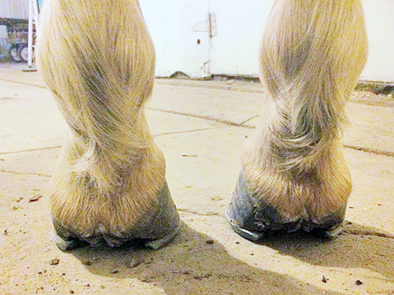
Recognizing And Managing The Club Foot In Horses Horse Journals

Hoof Evaluation Radiographs For The Farrier
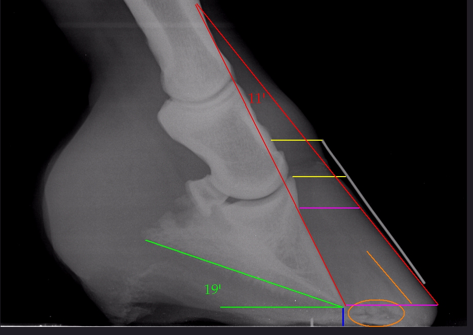
Understanding X Rays The Laminitis Site
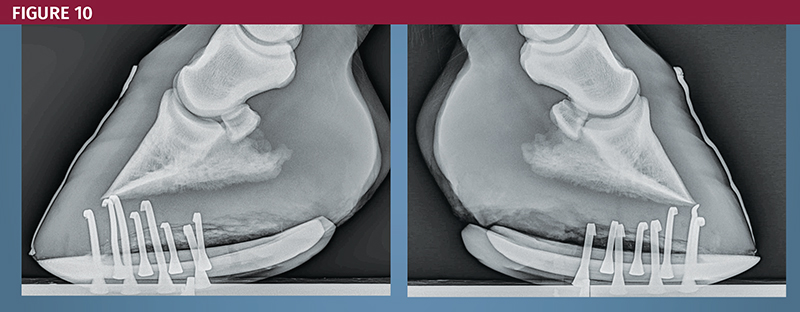
Recognizing And Managing The Club Foot In Horses Horse Journals
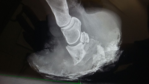
Rvc Equine Laminitis Facts And Research
Club Foot Or Not Barefoot Hoofcare

The Equine Documentalist Case 2 Lf Vet Interpretation High Heels Broken Forward Hpa Possible Laminal Pain Take Heels Down Farriery Interpretation Grade 1 Club Foot Caused By Contracture Of Flexor

No Foot No Horse Part 1 Hayes Equine Veterinary Services
